Successful dental cases require good digital scans. Today let’s discuss bad and good digital scans. We will also provide solutions, so you don’t have to call back patients for new scans from time to time.
With digital scans, dentists need to pay great attention to Interproximal contacts of the prepared teeth, because it is difficult to properly scan those areas. According to our experience, 80% of our digital dental cases with open margin is related to interproximal contacts.
1. Bad digital scans
![]()
Let’s look at the above example. The margin is subgingival. There is blood, and the gum tissue covers the margin. Therefore, the doctor could receive a crown that has open margin or fitting problem, because our lab technicians have to guess where the margins are, but this guess experience COULD be wrong.
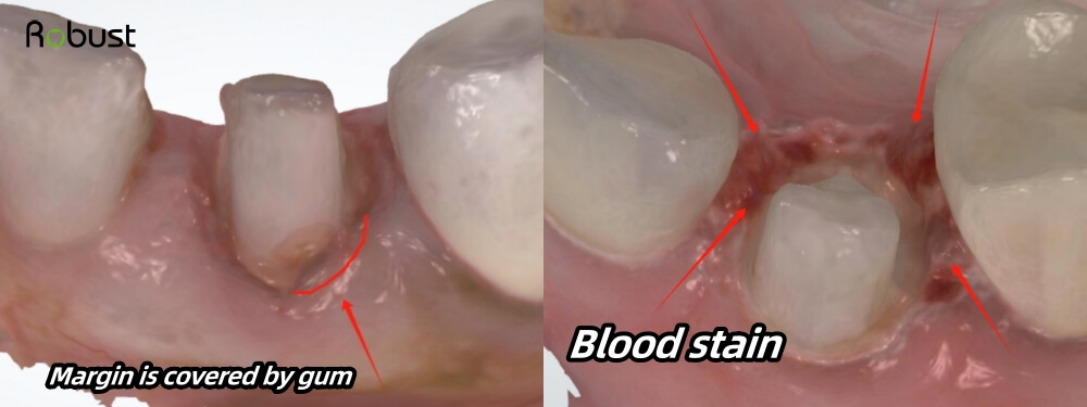
In the left example, the gum tissue is swelling and covering the margin. In the right side example, there is also a lot of blood stain. Those conditions would negatively influence our technicians’ judgement on margins.
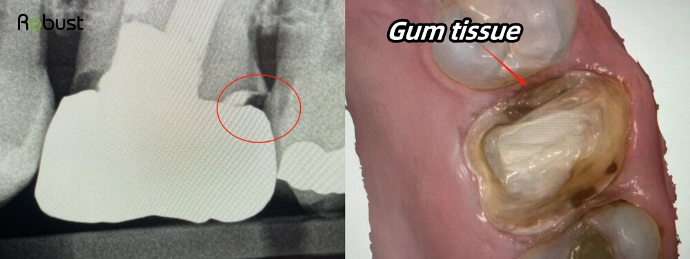
In this case, it seems the margins are clear, but if you look more closely, you will find a zigzag line on the mesial. It is because the mesial margin is covered by flesh (gum tissue). After a thin layer of gum tissue is cleaned with water several times, it becomes transparent. If this transparent gum tissue attached to the margin, it is hard to find out.
Normally, it is best to prepare teeth above gingival. Of course, we understand that dentists cannot prepare margins above the gum due to some special conditions. Therefore, if dentists have to prepare teeth subgingival, It is best to use retraction cords, which can help clearly separate gum tissue and tooth structure. Or if you don’t use retraction cords, please at least make sure you have totally cleaned blood and saliva, and no gum tissue is covering the margin.
2. Good digital scans
![]()
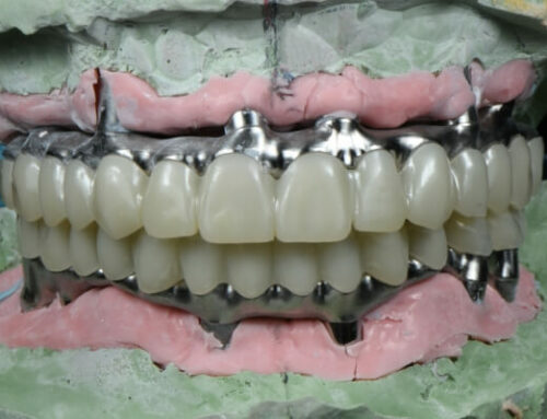
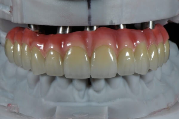
Let’s look at the above example. The margin is above the gum, so the gum tissue is not swelling. We can see all margins are clear. With this scan, our technicians can easily identify the margins and make crowns with perfect fitting.
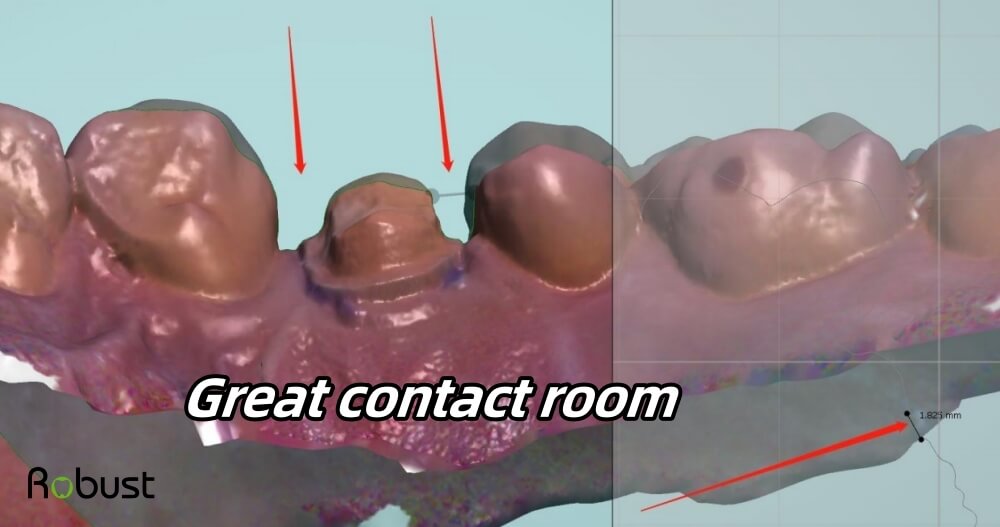
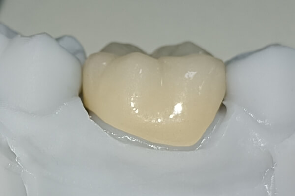
Second, each interproximal space is 1.825mm. Usually, the interproximal space should be between 1~1.5mm, so we can make a crown that is thick enough. Please note a crown should be about 0.8~1mm thick, in order to avoid breakage. With this interproximal space, dentists can also seat crowns with ease.
![]()
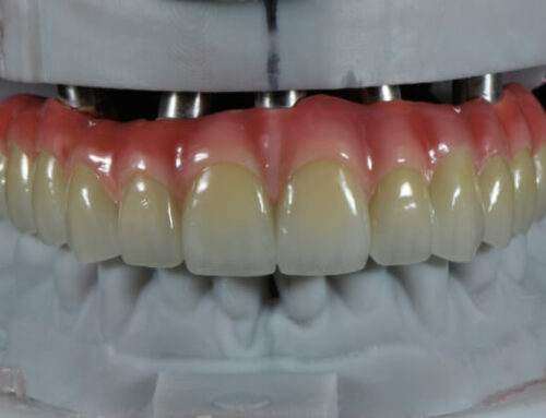
Third, the occlusal space is 2.621mm (the occlusal space normally should be between 1.5~2mm). This occlusal clearance is enough for us to make a crown with beautiful details. We can also make the final crown slightly out of occlusion, so the crown will not break easily due to strong biting forces.
![]()
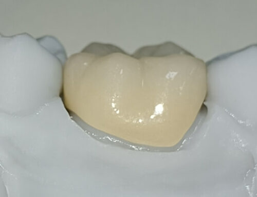
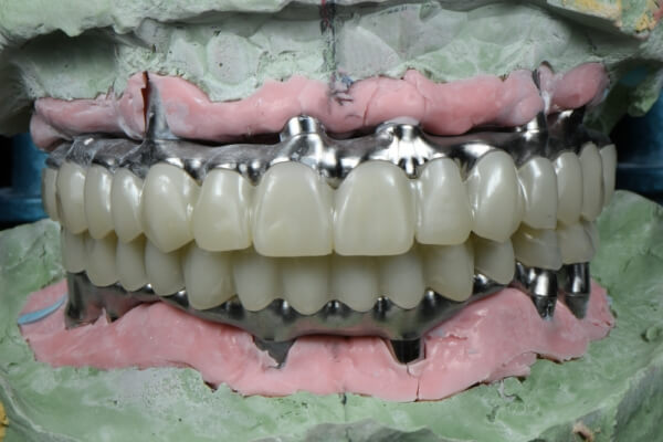
In this scan, the margin is subgingival. Yet, the doctor treated this case pretty well. Even though the gum tissue is swelling, the doctor separated gum from tooth structure (no gum covers margin). Also, she cleaned blood and saliva on the margin area.
3. Conclusion
In conclusion, good scans help dentists save time and cost. It will also make patients clinic visit easy and quick. If you follow these instructions and check your digital scans carefully before sending to us, we believe you can greatly decrease the remake rates.